


[051] Heartbeat pumping 2 (GB#103A02) | 基礎医学教育研究会(KIKKEN)Lab

●Invention of heart valve
The structure of the heart is as good as I thought what kind of genius thought it was. We are also impressed with clever piping, but it is particularly amazing to devise a way to pump blood with a simple mechanism. Before human beings invented hand pumps, they are putting practical use of valves completely (laugh).
—
Contents
- 1 ●Building a heart pump
- 2 ●Troublesome because it has a name on the right and left
- 3 ●Right and left have different flowing blood
- 4 ●Do not open the inlet and outlet valves at the same time
- 5 ●The valve opens and closes due to contraction and relaxation of the ventricle
- 6 ●Blood does not flow in myocardium during ejection period
- 7 ●Blood flowing in one heart beat
- 8 ●Both right and left blood flow volume is same
- 9 ●Heart stops moving even though myocardium is moving
- 10 ○Related articles
- 11 ○Referenced books
●Building a heart pump
There is an impression that the heart seen in the textbook has a rather complicated structure. But it combines two separate pumps of venous blood pump and arterial blood flow pump into one with appearance and movement, so either one can explain the basic mechanism of the workings of the heart . Simplifying, the basic configuration including the blood vessels before and after the heart, along the direction of blood flow,
It is arranged in the order of vein (blood vessel), atrium (heart), ventricle (heart), artery (blood vessel).
Although it is a single organ, each area has characteristics in its material and work. It is the atrium and the ventricle that make up the heart that repeat contraction and relaxation spontaneously. Since the heart is made up of striated muscles, it is good at repetition of quick and powerful contraction and relaxation.
On the other hand, veins and arteries, which are blood vessels, are made up of smooth muscles, which can not be quickly moved, but is suitable for maintaining sustained tension.
The central part that works as a pump is a ventricle, and the point is that there are “valves” which are doors that open only to one side at the entrance and exit.
Entrance door: atrioventricular valve, valve between atrium and ventricle
It opens only from the atrium to the ventricle.
Exit door: arterial valve, valve between ventricle and artery
It opens only from the ventricle to the artery.
●Troublesome because it has a name on the right and left
In English, the valves of the left and right arteries are called by their own unique names, and it seems that there is not much to mention the two together.
The right atrioventricular valve is a tricuspid valve.
The left atrioventricular valve is a mitral valve.
The right arterial valve is pulmonary valve.
The left arterial valve is the aortic valve.
Each blood vessel connected to the heart also has its own unique name.
The veins on the right are upper and lower big veins, vena cava.
The left vein is pulmonary vein.
The right artery is the pulmonary artery.
The left artery is the aorta.
As you can see, it seems like the components are confusing to beginners, as the parts are so many that they are named individually.
●Right and left have different flowing blood
 The contents of the flowing blood are different between the right pump and the left pump. The venous blood flowing through the right heart is the blood before it is supplemented with oxygen in the lung after it has traveled through the body. Arterial blood flowing through the heart on the left is fresh blood to be pumped into the body after replenishing oxygen in the lungs. It is a different route which does not mix with each other in the heart.
A pump through which arterial blood flows,
Pulmonary vein ⇒ left atrium ⇒ left ventricle ⇒ aorta
The pump through which venous blood flows,
Caval vein ⇒ right atrium ⇒ right ventricle ⇒ pulmonary artery
In other words, it is independent as a pump. Another difference is that the pressure for sending blood is very different between right and left. The left ventricle to be delivered to the aorta can reach as high as 120 mmHg to 130 mmHg, but the right ventricle that only sends blood to the lungs is enough for that one fifth. However, the two pumps repeat contraction and relaxation pulsation / beat at the same timing. So for now, do not distinguish right and left, think about the basic movement of the heart.
●Do not open the inlet and outlet valves at the same time
Since the movement of the heart itself is also a circulation type, it may start from anywhere, but usually it seems to start with “cardiac cycle” where the atrioventricular valve opens and sucks blood into the ventricle.
1) filling phase
Only the atrioventricular valve is opened, and the ventricle inhales blood from the atrium.
2) atrial contraction
The atrium pushes the blood into the ventricle only a little.
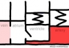
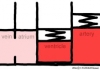
3) isovolumetric contraction
All the heart valves are closed and the pressure in the chamber increases.
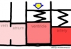
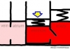
4) ejection phase
Only the arterial valve is opened, and blood is sent to the artery.
5) isovolumetric relaxation
All the heart valves close but the pressure in the ventricle goes down.
After that, only the atrioventricular valve was opened and returned to the initial filling phase, after that the same cycle was repeated about 70 times per minute (time of one beat was about 900 milliseconds), making it quiet all day If you do, repeat 100,000 times. Meanwhile, the atrioventricular valve and the arterial valve never open simultaneously.
●The valve opens and closes due to contraction and relaxation of the ventricle
 In the “filling period” when the atrioventricular valve opens, the pressure in the ventricle will instantaneously drop to around zero, and it seems to be in a form that draws blood from the atrium.
 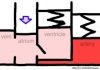 After that the atrium muscle contracts, followed by “atrial systole” which forces the blood a little bit into the ventricle. In the electrocardiogram, the P wave at the beginning of the cycle overlaps the onset of atrial contraction, so there is a way of explaining this as the beginning of the cardiac cycle. While the blood flows from the atrium to the ventricle, the arterial valve remains closed all the time. Since the opening and closing of the valve and the flow of the blood are not different between the filling period and the atrial systole, there are cases where this is sometimes completed as a filling period collectively without distinction.
 The QRS wave of the electrocardiogram (QRS complex) occurs immediately after the atrial systole, and the pressure rises as the ventricle begins to shrink suddenly, so the atrioventricular valve closes. This is the beginning of the “isovolumic systole”. The vibration that the atrioventricular valves hit each other at this time is the sound of the heart called the first heart sound.
Even when the atrioventricular valve is closed, the exit arterial valve does not open yet, so the ventricle is sealed. When the blood pressure on the artery side is 80 mmHg when it is an aorta (about one fifth if it is on the pulmonary artery side), the inside of the heart chamber just breathed still has a low pressure. 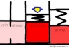 The arterial valve does not open until the ventricular muscle further constricts and the pressure in the ventricle overcomes the arterial blood pressure. During this short sealed state, the ventricular muscle contracts strongly while the ventricular blood does not enter and exit, and the volume does not change. It is an event of only 30 milliseconds in time.
 When the arterial valve opens, it becomes the “ejection period”, and the blood whose pressure has increased in the ventricle is expelled to the artery. As the contraction of the ventricular muscle becomes stronger, it pushes the blood into the strained artery as much as possible. Arterial blood pressure peaks beyond 120 mmHg in the left ventricle, then falls slowly as the blood of the artery is pushed out into the blood vessel ahead. On the other hand, the ventricular contraction force falls faster than the arterial pressure drop, and when the pressure reverses, the exit arterial valve closes at once and the ejection phase ends. At this time, the vibration that the arterial valves collide with each other is the second heart sound. (The whole will proceed with the right ventricle under the pressure of one fifth on the left.)
 When the arterial valve is closed, the atrioventricular valve remains closed, so that the ventricle is sealed again. This closed state is a phase in which the ventricular muscle relaxes, so it is called “isovolumic diastole”. The atrioventricular valve does not open until the contractile force of the ventricular muscle decreases sharply and the ventricular pressure falls below the atrium.
 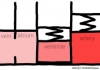 The isovolumic diastolic phase takes about twice as long as the isovolumic systole. Still it will be a momentary incident. When the atrioventricular valve opens, it returns to “filling period” again.
●Blood does not flow in myocardium during ejection period
From the isovolumic systole to the ejection phase, the myocardium contracts strongly. Blood vessels running in the myocardium are crushed by contraction of the myocardium, during which time the blood is swept out and does not flow into myocardium.  Instead, it is a mechanism that allows the heart muscle to relax by the blood pressure of the artery, from the isovolumic diastole to the point before the next isovolumic systole. Blood flowing while the myocardium is relaxing inflates the collapsed blood vessel. This phenomenon seems to be whether the shrieked ventricle regains the shape of the bag and plays a role in aspirating the blood from the atrium, so that when the air is again injected into the air doll where the air has gone out, I think, but how about you?
●Blood flowing in one heart beat
The blood volume in the ventricle is maximized just before the isovolumic systole, and about 120 ml is collected when it is at rest. It becomes the minimum immediately before the isovolumic diastolic phase through the ejection period, but it does not become empty, about 50 ml remains in the ventricle. Approximately 70 ml of that difference is the amount of blood that is pumped out in one heartbeat. However, the amount of blood flowing through the heart fluctuates slightly every time, and it varies greatly depending on how you move the body. When exercising, the heart rate also rises, but the stroke volume per one also increases.
If you consider it normally, there may be an image that the amount of blood remaining in the ventricle decreases by a small amount when the stroke volume is high, and the remaining blood volume increases when the stroke is small. However, the amount of blood remaining in the ventricle does not change basically regardless of whether the stroke volume increases or decreases. Generally, if you reduce the remaining amount in order to spit out a lot of blood, it will not return to its original capacity even if blood normally flows in the next filling period. Conversely, after decreasing the stroke volume by increasing the remaining amount, the ventricle expands to the extent that blood comes in normally. If the stroke volume is reduced in the same way, the ventricles further expands.

 The heart has an automatic mechanism that does not cause such inconvenience originally. When the myocardium is stretched long, it tries to return to the base stronger. It is a property called the Starling’s law of the heart. Therefore, when heart inhales much blood, there is a nature that the heart automatically expels a lot. If you inhale more, give out more, less as you inhale, give less. So basically the amount remaining in the ventricle remains unchanged. When the stroke volume increases to the maximum, it becomes about twice as much as at rest. Even if the stroke volume doubled, 50 ml remained originally, so it is 50 + 70 × 2 = 190 ml and the ventricular bulge is increased by up to 60%.
●Both right and left blood flow volume is same
Blood leaving the heart passes through the artery around the destination and takes the veins back and comes back. Blood leaving the left ventricle is arterial blood, goes through the systemic circulation circulating in the body, becomes venous blood and returns to the right atrium. Venous blood comes out of the right ventricle and is pulmonary circulation (pulmonary circulation) which is sent to the lungs and becomes arterial blood and returns to the left atrium. The blood volume of the pulmonary circulation is much less than the blood volume of the systemic circulation and the pressure of the pulmonary circulation is only about one fifth of the systemic circulation, so it may be thought whether there is a difference in stroke volume, The amount of flowing blood is exactly the same for both the right heart and the left heart. If stroke volume is not the same as right and left, circulation will be broken. Since the pressure for sending out blood has a large difference between right and left, the timing of opening and closing the valve seems to be slightly shifted right and left in reality. However, if you are not an expert, there is nothing to worry about.
●Heart stops moving even though myocardium is moving
 It is said that the heart stops when you die. Of course the heart is stopped when it dies. But, suddenly, even if “the heart has stopped”, the heart itself is not necessarily dead yet. To work as a pump, at least the ventricles inhale the blood and spit out, that much movement and time to allow the blood to flow. The ventricular streaks must repeatedly contract and relax greatly all at once as explained above. Unfortunately, even if it shrinks in a convulsive state in a small manner like ventricular fibrillation, the valve does not open and it is only wasteful activity. Even if the heart is moving, if it can not work as a pump, it is the same as the heart is stopped. It is this kind of “moving cardiac arrest” that can be helped by using AED in various facilities, that is, automatic external defibrillator as soon as possible.
○Related articles
◆[041] 心筋線維 myocardial fiber ![]()
◆[016] 血液循環 blood circulation ![]()
◆[001] 心臓のポンプ機能 heartbeat pumping ![]()
◆[008] 弁の働き valve in the thin‐walled vessel ![]()
◆[033] 平滑筋の収縮 smooth muscle contraction ![]()
◆[009] 筋収縮の伸縮幅 the range of muscular contraction ![]()
◆[035] 骨格筋収縮の張力 tension of the skeletal muscle contraction ![]()
○Referenced books
・カラー版 ボロン ブールペープ 「生理学」, 西村書店
・カラー図解 人体の正常構造と機能 全10巻縮刷版,坂井 建雄,日本医事新報社
・人体機能生理学,杉 晴夫,南江堂
・トートラ人体解剖生理学 原書8版,丸善
・柔道整復学校協会編「生理学」,南江堂
・東洋療法学校協会編「生理学」,医歯薬出版株式会社
rev.20160720,rev.20161022
KISO-IGAKU-KYOIKU-KENKYUKAI(KIKKEN)