


[014] Aaccommodation of the eye (GB#116G03) | 基礎医学教育研究会(KIKKEN)Lab

● When looking at closer things the ciliary muscle contracts and the crystalline body loosens
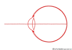
When viewing the closest thing, when seeing the distant thing, junior high school students also learn that the crystalline lens in the eye is adjusting the focus. It is a reaction called accommodation. The point is that the ciliary muscles around the crystalline lens contract when viewing near. Then the lens thickens and the focus of light comes to the retina. When the lens becomes thick, the focal length becomes short. The logic is simple. But mostly, by looking at the explanatory diagram attached next to it, how many people can be convinced by this mechanism smoothly?
—
Contents
●Which shrinks when the ciliary body contracts?
In the figure when viewing nearby objects it is good that the crystalline lens is thick. But, in many figures, the muscles that are supposed to be pulling from the side are not shrinking, do they seem to be stretching rather? Or, there is a figure that only the crystalline lens is swollen for some reason. Even if you look at any picture, it is difficult to understand this process of construction so badly.
Why is this happening? I doubt that those who have drawn explanatory diagrams may not really understand it. Of course, the authors of the explanation should be understood because they are experts, but maybe they have not properly communicated them to the illustrator.
● The lens is pulled from the surroundings like a trampoline
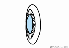
The crystalline lens of the eye looks like a globular elastic gelatin. Fibers called the ciliary body (Zinule of Zinn) are pulling around it. The ciliary body that goes around the outside pulls the fiber. The lens is always pulled from the surroundings. So the basic shape of the crystalline lens is somewhat flattened. The figure being pulled with a string is just like a children’s trampoline. However, although the ciliary body is in the frame position around the trampoline, it is not that stiff, and in fact the ciliary body itself is not pulling the ciliary body.
(But in fact, most of the explanation is confusing because the ciliary body draws the lens like pulling it.)
Then where does the pulling force of the ciliary body be from? Is it a ciliary muscle? That would be strange. The ciliary muscle is certainly a muscle in the ciliary body, but it should be explained that when the ciliary muscle contracts the crystalline lens becomes thicker. Apart from the ciliary muscle, there is another force pulling the ciliary body itself outward. The ciliary body is strongly bound to the sclera of the eyeball. Water is constantly injected inside the eyeball, and it keeps its round shape with a slight pressure but inflated steadily to the outside, intraocular pressure is applied. It is nothing but this intraocular pressure that is pulling the ciliary body. Let’s inflate the balloons in a purse bag fully. It is more likely to be a water balloon. The bag will swell round and the mouth of the purse string will spread. That’s it.
● When viewing near the ciliary body ring becomes smaller
The ciliary muscle is the smooth muscle surrounding the crystalline lens in the ciliary body. It seems to be a double structure of annular fiber (Muller muscle) running in a ring shape and radially running muscle (radial fiber and meridian fiber, Brucke muscle) orthogonally to it. However, let’s consider the ring-shaped fiber for the time being. What will happen if this shrinks? The rings surrounding the crystalline lens become smaller. Then the power pulling the lens outwardly weakens and the lens will inflate with its own elastic force.
Then what is the radially running muscle at this time? If it is a model where the ciliary body stretches when looking at near, this stretching will be radial fibers. However, radial muscles are also smooth muscle like annular fibers and should contract with parasympathetic acetylcholine. It would not seem that one is contracting and the other orthogonal is relaxing and doing something complicated. If so, it should be written in textbooks. Indeed, in pupils, it is written in most textbooks that the sphincter pupillae muscles and the dilator pupillae muscles are orthogonal and controlled by different nerves. As for the ciliary body it is only written that it contracts with the parasympathetic nerve, so in short it may be considered that both the annular fiber and the radial fiber shrink at the same time. What will happen then? The ciliary muscles will pull the surrounding sclera and reduce the ring. It’s like tightening the mouth of a drawstring bag that has inflated a balloon.
● When seeing near, will intraocular pressure go up?
What is inferred from now is that the eyeball will be a little smaller when looking at the nearby eye or the volume of the eyeball (vitreous humor) will be slightly smaller. However, the colloidal vitreous body is contained in the bag and does not mix with the aqueous humor, nor does it leak out from the side of the ciliary body. Then, it can be expected that the intraocular pressure rises slightly. How is it actually? Although experts will understand naturally, there is no information at least on the net. However, it seems that the person with myopia suffers slightly higher intraocular pressure. However, this will not be related to the movement of the ciliary muscles. It is a bit difficult to imagine that the ciliary muscle is kept taut all the time so that the intraocular pressure keeps rising all the way. It is because you can adjust to the normal range in that state. Conversely, if the intraocular pressure is high, the power to pull the crystalline lens becomes large, so the crystalline lens becomes thin and may worry about hyperopia rather than myopia. However, there is no such description. Apart from that, from long ago, people who were originally intensified in intraocular pressure should have been told to avoid nearsides, and there seems to be a situation in which the circumstances explained this time are in the background.
If you keep looking at the nearby place, your eyes get tired, and on the contrary it will be easier to see far away. Is not it related to this?
※ This animation highlights the parts of the ciliary flat (pars plana ciliaris) and saw-like rim (ora serrata) to float up due to ciliary muscle contraction, but in fact it is different. The longitudinal muscles are in a positional relationship such that the ciliary flat portion is pulled up toward the base of the iris (the iris origin). Therefore, when it contracts it may be like movement to elevate the whole ciliary body as much as squeezed by the cricoid. It seems that the eye itself is not deformable as much as possible. Nevertheless, it might be a movement that pulls up the saw-like edge and squeezes the inner surface of the eyeball.
● Referenced sites
* Accurately, “intraocular pressure” is measured from the outside of the eyeball, which is different from the original intraocular pressure, but generally it works in conjunction and the values seem to be about the same level. The normal value of intraocular pressure is set to 10 – 21 mmHg.
※ When the ciliary muscle contracts, a hole (Schlemm’s canal) for discharging the aqueous humor at the immediate side opens, and the eye pressure decreases.
* Actually, some people are troubled by being caught at the ciliary muscle.
⇒ Will the lens thicken as the ciliary body contracts? 【Link broken】
⇒ ⇒ Why does the tin zona become tense when the ciliary body relaxes?
· The science teacher is observing the chin band with pig’s eyeball [Because grotesquate pictures will come out, people who do not like it may be better to avoid seeing it]
· To apply such time and effort solely for eye accommodation adjustment. ⇒2004 Iwate Prefectural Education Workshop Presentation Material (High School Biology I)(Iwate Prefectural Comprehensive Education Center) ※ Please be aware that this is a bit more grotesque photos. Is it the same author as the above blog?
· Fun cartoon is also introduced. ⇒ Lesson 4 – 01 (p 8) Basic adjustment is “far” (Moe et! High school student I · II)
· A very detailed explanation by experts is done. ⇒The new concept of accommodation (Dynamic biomechanical model)
2015-06-25 Addition
○ Related articles
◆[020] 対光反射 pupillary light reflex ![]()
◆[044] 伸張反射 stretch reflex ![]()
◆[033] 平滑筋の収縮 smooth muscle contraction ![]()
◆[031] 興奮伝導 conduction of excitation ![]()
○ Referenced books
・カラー図解 人体の正常構造と機能 全10巻縮刷版,坂井 建雄,日本医事新報社
・人体機能生理学,杉 晴夫,南江堂
・トートラ人体解剖生理学 原書8版,丸善
・イラスト解剖学,松村 讓兒,中外医学社
・柔道整復学校協会編「生理学」,南江堂
・東洋療法学校協会編「生理学」,医歯薬出版株式会社
rev.20150625, rev.20170211, rev.20170503.
KISO-IGAKU-KYOIKU-KENKYUKAI(KIKKEN)