


[049] A neural circuit for voluntary movement (GB#114D08) | 基礎医学教育研究会(KIKKEN)Lab

● A comprehensive project involving brains, muscles and senses
In order to manipulate our body at will, a large amount of signals circulate around the brains. The command sent to the muscles is the result signal. In addition, unless confirmation of the effect of the executed motion and correction of the signal are made, it is not close to the movement as desired. The execution of voluntary movements is a big comprehensive project that also incorporates afferent signals from the periphery.
–
—
Contents
- 1 Organization of voluntary movement project
- 2 ● Two loops of basal ganglia and cerebellum
- 3 ● Two flows to perform movement
- 4 ● Check actual movement
- 5 ● Confirm actual feel
- 6 ● Choose an movement that fits your intention
- 7 ● “Extrapyramidal disorder” is a disorder of the extrapyramidal pathway?
- 8 ○ Referenced sites
- 9 ○ Related articles
- 10 ○ Referenced books
Organization of voluntary movement project
Movement of the face and limbs made by our own intention is voluntary movement. Sometimes there are casual movements that are not conscious of ourself, but regardless of whether we are aware or not, the basic mechanism of execution is the same.
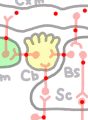
At least the following elements are involved in voluntary movement projects. (Abbreviations are effective only in this site. In addition, because the connection status of information is given priority in this chart, placement does not match the anatomical positional relation in some places. The relationship between the right and the left is also neglected here.)
Cxm: Cerebral Cortex (motor area).
Cxs: Cerebral Cortex (sensory area).
BG: Basal Ganglia.
Thm: Thalamus (motor nuculei).
Ths: Thalamus (sensory nuculei).
Cb: Cerebellum.
Bs: Brain Stem, Brainstem / Sc: Spinal Cord.
Ms: Skeletal Muscle.
S: Sensory organ.
* As a neural circuit for voluntary movement, the boundary between the brain stem and the spinal cord is not so clear in fact. Especially in the medulla, although there is a difference in the level of function, it seems to be an extension of the spinal cord. Also, the action of medulla oblongata and pons seems not to be clearly distinct as that form. So, here we are batching the brainstem and the spinal cord on purpose to make the story as simple as possible.
● Two loops of basal ganglia and cerebellum
A terribly rough explanation (you can only do a rough explanation on this site), in the brains that are in the skull (cranium), two large loops are spinning around for a voluntary movement.
【Loop of basal ganglia】
 · Cerebral cortex (Cxm) in the motor system ⇒ basal ganglia (BG) ⇒ thalamus in the motor system (Thm) ⇒ A loop returning again to the cortex of the motor system.
This loop organizes the goals and projects of movement according to the motive of movement and narrows down the optimal movement from various repertoires of movement.
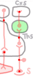
【Cerebellar loop】
 · Cerebral cortex of the motor system ⇒ Brain stem (Bs) ⇒ Cerebellum (Cb) ⇒ A thalamus of the motor system ⇒ A loop returning again to the cortex of the motor system.
While executing movement, correct errors of motor commands to muscles and preserve optimal correction pattern.
※ The thalamus (Th) looks like a united chunk of left and right respectively. But, for example, in terms of logistics, it is like a big wholesale district where large sections of goods are handed over at different times, with divisions divided by department. The thalamus of the motor system (Thm) and the thalamus of the sensory system (Ths) are separated, and basically the signal of the basal ganglia and the signal of the cerebellum flow in separate sections, even in the thalamus of the motor system.
● Two flows to perform movement
The cerebral cortex (Cxm) creates a motor program based on the information of the cerebral basal ganglia loop and decomposes it into contraction and relaxation motor command signals for each muscle or group of a few muscles and reorganizes it. It transmits its decomposed / rearranged signal to the brainstem (Bs) and the spinal cord (Sc) through the efferent’s descending tract. Motor neurons in the brain stem and spinal cord exert peripheral nerves called cranial nerves and spinal nerves and execute the activities of the respective muscles according to the signals. Neurons not going out of the brain or spinal cord are sometimes called roughly “motor neurons” that relay the motor system. Therefore, in order to clarify the position, neurons that directly move the muscles by peripheral nerve fibers are also called “lower” motor neurons. There are two major routes on the descending path from the cortex to the spinal cord.
【Pyramidal tract】
This is a descending route that carries a signal of motor command without interruption until the neural fibers of cerebral cortex neurons reach the brainstem or spinal cord lower motor neuron. Routes that extend to the spinal cord are called corticospinal tract by arranging the departure place and destination in an anatomical practice, but since most go through the pyramidal decussation of medulla oblongata, it is also called pyramidal tract.
Since the brainstem is above pyramidal decussation, the fibers reaching the lower motor neurons such as the facial nerve in the brainstem from the cerebral cortex do not go through pyramidal decussation. However, as a relationship to the lower motor neuron, it seems to be the same as the corticospinal tract, so this is also often treated as “pyramidal tract” functionally as a whole. We do not distinguish it even in this chart. When it is necessary to distinguish it from the corticospinal tract, say cortinuclear tract. In this case, “nuclear” refers to a mass of lower motor neurons in the brain stem, such as a facial nucleus. The corticobulbar tract is one of the cortical nuclear tracts relaying in the medulla oblongata, but it is also used as a synonym for the cortical nuclear tract.
![]() The region of the cerebral cortex that exits the pyramidal tract forms a special area called the “primary motor area”.
The region of the cerebral cortex that exits the pyramidal tract forms a special area called the “primary motor area”.
[Extrapyramidal tract]
Extrapyramidal tract represents the meaning of “not a pyramidal tract” among descending motor paths of the spinal cord. Certainly the pyramidal decussation is out. But it does not matter that it does not go through pyramidal decussation, the important thing is that there is a relay point in the brainstem. In other words, after the signal exits the cerebral cortex, it changes the neuron via the synapse at least once in the brain stem, and the nerve fiber that originates from the brain stem reaches the lower motor neuron.
Specifically, these routes include cortex ⇒ red nucleus ⇒ spinal cord route with the nucleus ruber of the midbrain as a relay point, or a reticular formation spreading from the top to the bottom in the brainstem, cortex ⇒ reticular formation ⇒ spinal cord route, or cortex ⇒ reticular formation ⇒ medullary motor nucleus root.
The fact that there is a relay point means that before the lower motor neuron is reached, signals are divided to other places, mixed with signals from others, a bit of reworking of signals of movement comes in. Nerve fibers running from the cerebral cortex to each relay point, including the primary motor cortex, come from a wider range of cortical areas. Anatomically, as “extrapyramidal tract” in which fibers are bundled, it refers to the red nucleus spinal cord tract leading from the relay point of the brain stem to the spinal cord and the reticulate spinal cord tract.
In summary, the motor command that reaches each lower motor neuron from the cerebrum is carried in the direct route (pyramidal tract) from the cerebral cortex and the indirect route (extrapyramidal tract) relayed by the brain stem. For the direct route, the motor command considered by the cerebral cortex is transmitted as it is, and the indirect route gives a slightly revised motor command in the brain stem. A signal that captures and coordinates both commands and in situ information from the muscles and tendons via the spinal reflex circuitry around the lower motor neuron causes actual skeletal muscle movement through the lower motor neuron.
● Check actual movement
There is no guarantee as to whether or not it is the movement you want, even if you put effort into the muscle according to the motor command. Therefore, the motor system mobilizes various sensory functions all at once and checks the results of the movement instantaneously. The most fundamental thing is information on states such as muscles, tendons and joints, called the deep sense (proprioception). Familiar muscle spindles and tendon organs and the like instantly fine-tune the activity of lower motor neurons with spinal reflex while raising information to the brain one by one through ascending conduction pathways.
Where to go in the brain, the important destination in motor control is the cerebellum (Cb). To the cerebellum, a copy of the motor command going from the cerebral cortex to the lower motor neuron is input via the brainstem with another route. It is said that in the cerebellum, cortical motor command and actual body movements are checked. The greater the error range of the result, the stronger signal is returned to the cerebral cortex making the motor program through the thalamus. The cerebral cortex corrects the motor program according to the signal from the cerebellum, and transmits the motor command again.
Every time this loop is turned during an action, the error becomes small and it approaches the target of movement. With instantaneous movement, this correction will not make it in time. Even if it does not work in a single operation, the repetition of the operation many times will reduce the range of correction. By repeatedly executing various motor programs, each modification pattern seems to accumulate. This error correction loop is also a circuit of motor learning at the same time.
Frequently cited in motor learning is driving a bicycle. There will not be so many people who can ride a bicycle for the first time and run soon. Perhaps everyone will be able to run very well at first. However, with repeated excercises, almost anyone will be stable. Once you can get on, it is normal that even if you have not been riding a bicycle after that, even if you have forgotten to ride, it will not take long to get on again. The invention of a bicycle is the last thing in human history, and although the “bicycle riding program” is not prepared in the human brain in advance, the pattern of the motor program acquired by practicing is properly It means that it is memorized. Even if you forget the knowledge of the textbook you studied at junior high school, even if you forget it, the memory of the motor program thus acquired lasts for a long while without using it. This is an important point for living by using the body well while pulling out accumulated movement patterns according to various situations. It goes without saying, however, that special efforts are required to maintain the best performance just like the athletes.
Since the important checkpoint of motor learning is the cerebellum, when the cerebellum is affected by the lesion, the data of the motor program accumulated so far can not be used. It is essentially the same as having the data disappeared. So you have to recreate the motor program a bit, but the checking function does not work so you can not fix it. When the cerebellum is affected, although it can roughly move the skeletal muscle, it becomes a motor dysfunction “ataxia,” which is not effective in regulating force or direction. On the contrary, the work of the cerebellar loop is explained from the study of such a disorder.
● Confirm actual feel
 Besides motor equipment such as muscles and tendons, it is also important to check the results of movement by touching it with your eyes. Even if you hit the keyboard, the computer that the monitor disappeared would not be very useful even if it is moving. Even if you grab things, what will happen if you do not touch it? Let’s grab a rubber ball with a magic hand by remote control while looking at the screen of the personal computer. Even if it can be seen on the monitor, if the magic hand does not convey the feeling of touching the object, if you only operate slowly while watching the situation, you will skip the ball rather than grabbing it.
The stimulation of the skin generated by the actual movement is instantly sent to the cerebral cortex through the thalamus (Ths) of the sensory system. Therefore, it seems to work on confirming the motor program by exchanging information with the motor cortex as well as causing a sense of touch sensation in the sensory cortex (Cxs).
In a video game or the like, players “fight” with ultra-fast movement even without touch. However, it does not know how to be conscious, but it is established because it divides it into another world that was totally outraged. A mechanism that smoothly executes ordinary realistic voluntary movements is a system integrated with feedback (feedback) of information about various situations caused by exercise.
● Choose an movement that fits your intention
In the first place, you do not know where the motivation to do some movements, for example to ride a bicycle, or to take a certain posture comes from. However, if motivation is decided, it is not easy to say that the motor program will be decided. Even with an intentional movement, there is no single actual procedure. Every day in various situations, a new motor learning has been done, and a repertory of motor programs has accumulated in myriads. However, because there is only one body, only one of them can be executed at a time. For example, you go from your home to a friend ‘s house, there are various directions, and it is completely free to pick which one you choose, there is only one way to walk at a certain point.
Thinking about the weather at that time and other things to do, you will choose the way. Roughly speaking, it can be said that it is the loop that goes around the basal ganglia (GB) that judges the situation according to the intention and purpose of the motive and narrows down the program to be executed as it is concerning movement .
The reason is that the typical symptoms when the basal ganglia is affected by the lesion is that involuntary movement, in which a coherent movement does not stop arbitrarily and stops. From the name it seems like an “unsuccessful” movement but in fact the “uncontrollable” movement is closer. There are also some variations in involuntary movements, but it seems like a form of fighting trying to do some movement at the same time, regardless of intention, failing to narrow down the movement anyway. The output from the basal ganglia to the thalamus mainly works on inhibition. Involuntary movements can be thought of as causing weak activation of the cerebral cortex by suppressing this suppression to a level weaker than usual and not suppressing thalamic activity.
Meanwhile, there is a famous disease, Parkinson’s disease. This is also known as the disease of the basal ganglia, but this is characterized by the fact that the body solidifies and the movement is extremely small. At this time, suppression from the basal ganglia is too strong, suppressing thalamic activity. Therefore, cerebral cortex activity is also weak, it is thought that you can not choose motor program. With this disorder, if there is a specific instruction to the goal of movement, you can begin exercising relatively easily. This is why you proceed anyway if you choose to choose one of the paths to your friend’s house, but it seems that you alone are not resolved without being decided. The “resting tremor” that the fingertips will continue to move freely while still at rest is like a pieceita of hardship.
Basal ganglia was attracted attention due to obstruction of the motor function whose symptoms are easy to see. However, the basal ganglia as a whole has a signal loop that spans the entire frontal lobe. For that reason, it is thought that functions that do not appear as movement, such as thought functions, work similarly to movement.
● “Extrapyramidal disorder” is a disorder of the extrapyramidal pathway?
Movement disorders such as involuntary movements and restless tremor that appear in disorders of the basal ganglia were formerly called “extrapyramidal symptoms”. Even now, it may be understood by medical practice, but it is not used in national examination.In the era when the knowledge of neuroanatomy was incomplete, the mechanism of motor control was supposed to be “easy to understand” schema called a bimodal conception of the extrapyramidal system controlled by the basal ganglia via extrapyramidal pathway, in parallel with the cerebral cortex ⇒ cone pathway system of the pyramidal tract. Extrapyramidal symptoms are a remnant of that age.
The reason why this term is no longer used is that subsequent studies have shown that extrapyramidal tracts are also predominantly dominated by the cerebral cortex. In other words, it has been found that there are only a limited number of routes directly going from the basal ganglia to the extrapyramidal route, such as a part related to the mind unconscious walking pattern. In the end, it means that the actual condition of the “basal ganglia – extrapyramidal system” which had been supposed before has become disappointing. Although the anatomical “extrapyramidal tract” still exists, it is difficult to explain many “extrapyramidal symptoms” by the extrapyramidal lesions.
However, in textbooks that are not professional, these information are not fed back easily. “Extrapyramidal system” and “extrapyramidal symptoms” are still alive and confusion of terms and concepts will continue for a while in Japan.
○ Referenced sites
・Motor control, Wikipedia(2017)
・Motor coodination, Wikipedia(2017)
・Degree of freedom, Wikipedia(2017)
・マイナビニュース, 行動を決めるのには大脳基底核と前頭葉連合野が連携が重要 – 東京都医学研(2013年8月)
・小脳から大脳への出力形成メカニズムを解明|東京都医学総合研究所.(2014年10月)
・筧慎治(2001), 大脳小脳連関:小脳は大脳にどんな貢献をしているか -運動制御の観点から.
・小脳の学習と内部モデル (眼球運動を題材 に) ,川人光男, J-STAGE Journals- 日本神経回路学会,2001.
・運動麻痺と皮質網様体投射(2014)(リンクは張れないので検索してダウンロードしてください。)
・高草木薫(2009), 大脳基底核による運動の制御.
・平井宏明ほか(2012), 筋拮抗比の概念に基づくヒト歩行動作の運動要素分解.
・不随意運動, Wikipedia(2017)
○ Related articles
◆[044] 伸張反射 stretch reflex ![]()
◆[054] 気道と食道の切り替え switching of the airway and esophagus (GB#105A04)![]()
◆[040] シナプス伝達 neural signal transmission ![]()
◆[021] 活動電位 action potential ![]()
◆[031] 興奮伝導 conduction of excitation ![]()
◆[009] 筋収縮の伸縮幅 the range of muscular contraction ![]()
◆[035] 骨格筋収縮の張力 tension of the skeletal muscle contraction ![]()
○ Referenced books
・カラー版 ボロン ブールペープ 「生理学」, 西村書店
・カラー図解 人体の正常構造と機能 全10巻縮刷版,坂井 建雄,日本医事新報社
・トートラ人体解剖生理学 原書8版,丸善
・柔道整復学校協会編「生理学」,南江堂
・東洋療法学校協会編「生理学」,医歯薬出版株式会社
rev.20151224, 20151225, rev.20160725,rev.20170709,rev.20170714, rev.20171222, rev.20171224, rev.20171229.
KISO-IGAKU-KYOIKU-KENKYUKAI(KIKKEN)