[003] Diaphragmatic breathing (GB#104A01) | 基礎医学教育研究会(KIKKEN)Lab

● Lungs have no power to suck air
The lungs are made up of hundreds of millions of gigantic small bags “alveolus” that exchanges air and blood gas. The other space is occupied by a number of finely branched “bronchi” to allow air to pass through the coronary alveoli and a vascular network that circulates blood to the same extent.
In short, except for the blood vessels, the lungs are just made up of bags and tubes filled with air, not muscles. The lungs themselves have no movement power to suck or vomit air.
—
Contents
- 1 ● Lungs and containers are not stuck together
- 2 ● When breathing in, the diaphragm contracts
- 3 ●”Pleural space pressure is always negative pressure”
- 4 ● It is ridiculous when air enters the pleural space
- 5 ● Does the pleural space pressure not become positive pressure?
- 6 ○ Related articles
- 7 ○Referenced books
● Lungs and containers are not stuck together
The lung is packed in a container called “thoracic cavity” made of a solid cage called “chestthe thorax” and the bottom diaphragm. Because the thoracic cavity spreads and deflates with the help of the muscles, the lungs are inflated and deflated as a result of it. That does not mean that the lungs and the chest cavity are not stuck together. Rather, there is a slight gap so as not to stick.
This gap is called ‘pleural cavity’, and here is the important point.
* “Pleura” has two membranes, a thin “visceral pleura” wrapping the whole lung mass and a thin “wall side pleura” covering the inner wall of the rib cage. The gap between the two pleural membranes becomes the pleural space.
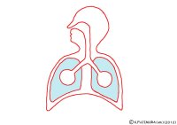
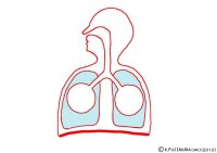
In the animation, the round bag connected to the bronchus is the whole lung of the alveolar mass, and the light blue part corresponds to the pleural cavity. The pleural space is actually filled with only a small amount of thin body fluid rather than the “space” containing air, and there is almost no gap. Thanks to this thin body fluid “serum”, the two pleural membranes do not stick together and the lung membrane slips well in the thoracic cavity.


● When breathing in, the diaphragm contracts
The diaphragm (thoracic diaphragm) is the wall that separates the thoracic cavity from the abdominal cavity. This wall comes out with muscles radially spreading fibers around the central connective tissue. It is a striated muscle, the structure and the moving mechanism are the same as the skeletal muscle.
When the chest cavity expands, the diaphragm may have an image to stretch. In fact, however, the diaphragm’s muscles contract when breathing. The diaphragm is stretched when the thoracic cavity becomes smaller, that is, when exhaling, and it is strongly pushed upward by the pressure from the abdominal cavity beneath it. When breathing in, the diaphragm contracts and pushes back the abdominal cavity downward. The chest cavity spreads. The breathing method which consciously utilizes only the force of this diaphragm is “abdominal breathing” (diaphragmatic breathing). This animation is an abdominal breath.
The diaphragm is a visceral organ, but the muscle is a skeletal muscle, and it is moved by a motor nerve like a limb. Even though all the nerves of the skeletal muscle around the diaphragm are coming out of the spinal cord of the buckwheat, the nerve that activates the diaphragm is somehow the phrenic nerve that emerges from above the neck. When severe cervical spinal injury occurs, the neck to paralyze down and the ribs can not move. However, if the phrenic nerve is not damaged, it can breathe by the diaphragm and will not suffocate.
**********
●”Pleural space pressure is always negative pressure”
This is a cliche in the National exam. Negative pressure is the power to suck. When sucking in juice with a straw, the inside of the mouth is negative pressure. In other words, as the pressure in this gap is lower than the atmospheric pressure, the lungs (alveoli) are pulled by the rib cage and spread due to the negative pressure. This means that the negative pressure of the pleural space is still maintained when the alveoli inhale the air, as well as when exhaling the air. Because, when you exhale, if the pressure in this gap becomes higher than the atmospheric pressure and it is positive pressure, the alveoli will collapse.
Once the collapsed alveoli do not spread easily. The textbook explains that it is the same as trying to pinch and spread the plastic bag whose inside was wet and crushed.
That’s why the alveoli are inflated by being constantly pulled by negative pressure so that they do not collapse even if they breathe out the air. Therefore, even if you exhale all the air exactly and try to make the lungs empty, about 1 liter is surely air remaining in the lungs. The amount of this air is named “residual volume”.
● It is ridiculous when air enters the pleural space
Both the alveoli and the pleura are very thin membranes. Since the pleural space is always negative pressure, as soon as there is a small hole in the wall of the lung, air comes out of the alveoli. The alveoli will collapse accordingly. Every time the chest cavity spreads due to breathing exercise the lungs collapse more and more, the alveoli can not breathe. It’s named “pneumothorax” and it seems not to be a particularly rare disease.
● Does the pleural space pressure not become positive pressure?
If it is always negative pressure, but “to atmospheric pressure”, this phrase will be accompanied by a provision that “When you are breathing in a natural way.” When stopping breathing and putting a lot of effort into the body, when inflating the balloon, the lung naturally exceeds the atmospheric pressure. Therefore, the pressure above the atmospheric pressure will be applied to the pleural space as well. But even at this time, the pressure in the pleural space should be lower than in the alveolus, if you compare the pressure of the alveoli with the pleural space. Otherwise the alveoli will collapse.
Negative pressure around the lung is important considering the alveolar blood circulation. When stopping breathing and tightening the thoracic cavity, the blood vessels in the lungs are also tightened. Meanwhile, gas exchange in the alveoli will have stopped. Conversely, the blood circulation of the lung is better if the pressure around the lungs is lowered as much as possible. If you take a deep breath and spread the chest cavity, it will be easier not only to get in and out of the air but also to exchange the blood.
⇒For those who would like to know more about the alveoli
⇒For those who would to know more about the pleural space
* In fact, commentary that can be recommended on the Internet is hard to find. Even on a proper site, be careful as there are clearly wrong content.
⇒ The students told me they do not understand the word “negative pressure”. Practical explanation is “to suck in”. As was also written above, in addition to the power to inhale the juice with a straw, the force when sucking in the garbage with a vacuum cleaner, the power when the lid of the bowl that went to stop opening, the udon noodles It is the power to suck in when the lap put on it is dented and it is attached to the juice.
It seems to be the most effective to have this negative pressure experienced in an experiment that inhalates a rubber balloon in your mouth and inflates it. However, since it is troubled if you inhale it wrongly, it makes me hesitate to make it all at once in lessons etc.
⇒Anatomical physiology 13 episodes “Trachea and lung”(Teacher Tama’s lesson)
○ Related articles
◆[015] Costal breathing![]()
◆[010] Alveolar ventilation and dead space ![]()
◆[050] Cellular respiration![]()
◆[011] Body acid-base reaction and carbon dioxide gas![]()
◆[023] Erythrocyte and hemoglobin![]()
◆[029] Oxyhemoglobin dissociation curve ![]()
◆[016] Blood circulation![]()
○Referenced books
・臓単―ギリシャ語・ラテン語 (語源から覚える解剖学英単語集 (内臓編))
・カラー図解 人体の正常構造と機能 全10巻縮刷版,坂井 建雄,日本医事新報社
・人体機能生理学,杉 晴夫,南江堂
・トートラ人体解剖生理学 原書8版,丸善
・イラスト解剖学,松村 讓兒,中外医学社
・柔道整復学校協会編「生理学」,南江堂
・東洋療法学校協会編「生理学」,医歯薬出版株式会社
rev.20150429,rev.20160403,rev.20170205, rev.20180714.
KISO-IGAKU-KYOIKU-KENKYUKAI(KIKKEN)


