[044] Stretch reflex (GB#114D04) | 基礎医学教育研究会(KIKKEN)Lab

● Reflex does not occur without nerves
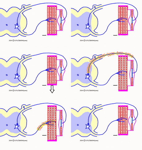
We can not usually be conscious, but skeletal muscles suddenly stretch and react to respond to contraction. However, the muscle cells themselves do not respond to pulling themselves. This is a “reflex” phenomenon through the nerve, which occurs when pulling the muscle, so it is called “stretch reflex”. Stretch reflex is the reflection with the simplest mechanism.
–
Contents
- 1 ● Initially muscle spindle stretching
- 2 ● There is a sensor body in the muscle spindle
- 3 ● Muscle spindle sensor to notify the difference in length
- 4 ● Skeletal muscle contracts when muscle spindle is pulled
- 5 ● The simplest mechanism reflex
- 6 ● What is stretch reflex for?
- 7 ● Stretch reflex works during exercise (?)
- 8 Misunderstanding of the role of stretch reflection?
- 9 ● Muscle spindle of iridescence
- 10 ○ Referenced sites / materials
- 11 ○ Related articles
- 12 ○ Referenced books
● Initially muscle spindle stretching
Basically, in a cluster of skeletal muscles, several elongated devices called “muscle spindles” of several millimeters in length are buried around the gaps of the muscle fibers. As the name suggests, it has a spindle shape with its center bulging in its entirety, and both ends are connected to the sheath of adjacent muscle fiber cells. Some of the muscle spindles near the ends of the muscles are connected to tendons on one side. This is an arrangement called “parallel relation” to the surrounding muscle fibers, so the muscle spindle also lengthens or shrinks as the whole muscle stretches and shrinks. And the muscle spindle acts as a stretching sensor which generates an excitation signal when lightly stretched. It is this muscle spindle that signals when a stretch is pulled.
● There is a sensor body in the muscle spindle
![]() The structure of the muscle spindle is a bit complicated. What is visible on the outside of the muscle spindle is a bag, in which several special muscle fibers run a lot narrower than the surrounding muscle fibers. Because it is a muscle fiber in the muscle spindle, it is called “intrafusal muscle fiber”. On the other hand, when referring to the muscle fibers occupying the major part of the muscle outside the muscle spindle, we call it “extrafusal muscle fiber” in the sense of the muscle fiber outside the muscle spindle. The central part of the intrafusal muscle fiber has a special structure, which seems to call this “equatorial region”, but this name is not listed in ordinary easy textbooks. However, since it is this part that is important with muscle spindle, we use it here. There is no property as a muscle in the equator part, and it has a certain length by elasticity. When the whole muscle spindle is stretched, it is this equator part that actually becomes longer. When the equator is stretched, an excitement signal is generated in the afferent nerve fibers (group Ia afferent fiber and group II afferent fiber) connected to it.
The structure of the muscle spindle is a bit complicated. What is visible on the outside of the muscle spindle is a bag, in which several special muscle fibers run a lot narrower than the surrounding muscle fibers. Because it is a muscle fiber in the muscle spindle, it is called “intrafusal muscle fiber”. On the other hand, when referring to the muscle fibers occupying the major part of the muscle outside the muscle spindle, we call it “extrafusal muscle fiber” in the sense of the muscle fiber outside the muscle spindle. The central part of the intrafusal muscle fiber has a special structure, which seems to call this “equatorial region”, but this name is not listed in ordinary easy textbooks. However, since it is this part that is important with muscle spindle, we use it here. There is no property as a muscle in the equator part, and it has a certain length by elasticity. When the whole muscle spindle is stretched, it is this equator part that actually becomes longer. When the equator is stretched, an excitement signal is generated in the afferent nerve fibers (group Ia afferent fiber and group II afferent fiber) connected to it.
● Muscle spindle sensor to notify the difference in length
The muscle spindle (to be exact, the afferent fiber surrounding the equator of intrafusal muscle fibers) is a sensor that generates an excitement signal when stretched. Since some force should be applied to stretch it, it is easy to misunderstand that it is a sensor of enlarging force. However, it is the extrafusal muscle fibers surrounding it and the tendon connected to it, to take on the strength of the muscle at that time. The muscle spindle can not measure the strength of the force, as the force of the extrasys is strong or weak, but the equator reacts as soon as it is stretched anyway, soon it reacts. Moreover, the muscle spindle was excited only when the equator part extended, and it is quiet when shrinking. It is to inform you that the work of the muscle spindle has extended for a while from a certain length.
Is there a sensor to measure the force of the muscle, it is not in the line. It is said that “Golgi tendon organ (= tendon spindle: although this expression does not appear in English)” in the tendon “connected in series” to the muscle is measured the strength of the muscle.
● Skeletal muscle contracts when muscle spindle is pulled
 If the muscle is pulled towards both ends for some reason, the muscle spindle, along with the extrafusal muscle fiber, that is, the equatorial part of the intrafusal muscle fiber in it is also stretched. Then, an excitement signal comes out from the equator and travels along the afferent nerve, and as soon as it enters the spinal cord, it immediately transmits an excitement signal to the efferent neuron. This efferent neuron is an Aα motor neuron (alpha motorneuron) that dominates the extrafusal muscle fiber pulled initially. When the Aα motor neuron is excited, extrinsic muscle fibers under its control undergo concurrent contraction through transmission of a neuromuscular junction.
It is easy to observe the phenomenon of stretch reflex. Tests such as patellar tendon reflexes or Achilles reflexes, which are called deep tendon reflexes in the clinical setting, are probably called because they are generated by tapping the tendon probably, but in reality they are all tests to verify stretch reflexes. By striking a tendon, the muscle spindle responds to it because it is very slightly stretched really truly. This can be tried by anyone as a simple means of checking whether there is no abnormality in the whole neuromuscular element directly involved in the specific reflex circuit or whether the central nervous system thereon is abnormal.
● The simplest mechanism reflex
Stretch reflex is the simplest mechanism in all the reflexes occurring in the human body. There are only two neural elements between the muscle spindle that generates the trigger signal of reflection and the extrafusal muscle cells reacting to that signal. Only afferent nerves that carry signals to the spinal cord and motor neurons that carry signals from the spinal cord to extrafusal muscle cells. In addition, in the spinal cord, afferent nerves are excitatory synaptic contacts directly to this motor neuron. ![]() Such reflex by direct communication between the input neuron and the output neuron is called monosynaptic reflex. Among all the reflex, the monosynapse reflex is only stretch reflex. (In the reflex circuit other than the stretch reflex, the afferent nerve signal finally reaches the efferent nerve after being relayed by several neurons in the spinal cord.) Furthermore, both afferent fibers and efferent fibers involved in stretch reflex are the thickest myelinated nerve fibers in the body. Reflex phenomenon is characterized by the fact that it takes a certain amount of time because the signal goes to the center and returns to the muscles. Among them, stretch reflex is also the shortest “latency”, the time from stimulation to reaction appearance. It is a human patellar tendon reflex with a long distance to the center, probably around 1 / 20th of a second.
Such reflex by direct communication between the input neuron and the output neuron is called monosynaptic reflex. Among all the reflex, the monosynapse reflex is only stretch reflex. (In the reflex circuit other than the stretch reflex, the afferent nerve signal finally reaches the efferent nerve after being relayed by several neurons in the spinal cord.) Furthermore, both afferent fibers and efferent fibers involved in stretch reflex are the thickest myelinated nerve fibers in the body. Reflex phenomenon is characterized by the fact that it takes a certain amount of time because the signal goes to the center and returns to the muscles. Among them, stretch reflex is also the shortest “latency”, the time from stimulation to reaction appearance. It is a human patellar tendon reflex with a long distance to the center, probably around 1 / 20th of a second.
There seems to be a difference depending on the type of muscle, but in one muscle dozens of muscle spindles are scattered and several afferent nerve fibers are also from each. There are countless motor neurons in the spinal cord, and the number of skeletal muscle fibers is many times that number. So actually, it is not as simple as drawing, but it is amazingly simple as a mechanism of a neural circuit of a vertebrate such as a human being by simply having dozens of identical sets together.
● What is stretch reflex for?
Stretch reflex is a simple neural circuit. So if you arrange the conditions as well, it will always appear in normal cases, it is an important point used for testing the nervous system. If there is movement disorder such as no force, deep tendon reflex disappears, there is a strong possibility of peripheral nerve disorder rather than disorder of the upper centers. Otherwise, if deep tendon reflex is elevated due to movement disorder, there is a problem in the upper centers rather than peripheral nerves. However, the stretch reflex neural circuit is not prepared for “nervous system testing”. It is not easy to explain briefly the original role of stretch reflex.
First of all, in this healthy state where this reflex works properly, we can not understand its ‘blessing’ at all. The flexion reflex where the fingers unintentionally come in contact with the needle and the light reflex squeezing the pupil when the light hits the eyes can be easily understood when the effect is working normally. In comparison, the tendon reflexes will not be visible on the surface as soon as they become conscious, so they do not understand as much as getting irritated.
Then, what if the stretch reflecx does not work well? When the stretch reflex is disappearing due to peripheral nerve injury, almost the entire peripheral nerve is damaged. Therefore, it is impossible to ascertain the effect of afferent fiber alone. There are diseases in which motor neurons are selectively impaired, such as amyotrophic lateral sclerosis (ALS). In the case of such peripheral motor neuron disorder, of course, there is motion paralysis and the extension reflection disappears. However, I have never heard of diseases in which only afferent fibers, in particular afferent fibers from the muscle spindle, are selectively impaired. (There is no confirmation, but it is sure to be listed in the textbook, if any.)
● Stretch reflex works during exercise (?)
Tendon reflexes can be observed only when the muscles are out of force and the joint is loosened. For this reason stretching reflections create a misunderstanding that it works only when the muscles are relaxed. When dragged by this, it means that you can not understand why there is such reflex because of what.
Although it is omitted in this rudimentary animation this time, the actual muscle spindle has a mechanism in which the length of the intracave muscle fiber is adjusted in real time in synchronization with the change in the length of the muscle accompanying voluntary movement. Even if the muscle stretches with voluntary movements, the length of the equator is constant and not stretched unnecessarily, while the muscle stretches at least as directed by the brain.Also, it seems that the muscle fibers are regulating every moment so that the muscle fibers in the spindle do not sag even during the movement in which the muscle shortens with voluntary movements. (The explanation of this mechanism is omitted here because it becomes complicated, but if you do not know the mechanism, the following story makes no sense, so if you want to know, please try searching “Adjustment of intrafusal muscle fibers”. )
What is going to happen is that the muscle spindle constantly monitors the length change of the muscle according to the brain command, even during movement, especially when shortening the muscle and moving the joint. When you try to lift something with your strength, if you can lift as you expect, the muscle spindle does not work particularly conspicuously (it may be keeping a constant weak signal). But if the baggage is heavier than you anticipated, that is, if you do not lift with the power that the brain commanded this power, the skeletal muscles will not shrink as much as the brain did. On the other hand, only the muscle spindle receiving another nerve command is contracting to the pre-calculated length. If the muscle spindle as a whole is going to shrink, if it does not shrink according to the calculation, if it is said that the discrepancy extends, the equator part of the intrafusion muscle fiber is stretched. Then, a strong excitement signal comes out from the muscle spindle, and as a result, it will contract the muscle a little more strongly.
Of course, the stretch reflection circuit of the muscle spindle also works to adjust the length of the muscles when maintaining a certain posture. However, under more normal circumstances, is not it supposed to be working as an automatic power booster to advance voluntary movements as planned (ceokikken ‘s guess). What is more important than closer to the workplace is an important point. It’s a neural reflex, so even though it is fast, it will be delayed by about 1/10th of a second, but it is faster and more practical all the time than it is to deal with it as far as reaching the brain.
* It is not clear yet, but in the academic, unexpectedly the explanation that “extensional reflex is being suppressed during voluntary exercise” is also impressive. In exercises where throwing objects at least vigorously, running or jumping, the muscle length quickly shortens, the adjustment by muscle spindle reflexes will not make it in time, so it is more reasonable to have stretch reflections disabled in advance maybe. Of course, stretching reflections should be suppressed in the stretching direction during voluntary exercise. Everyone has studied at a great pace, so in a scene where walking, grasping or lifting ordinarily, you want to think that stretch reflection works effectively in the background, do not you think? (Modified 2018-03-29)
Misunderstanding of the role of stretch reflection?
Mostly, it is hardly written in any textbook about the role of stretch reflex for some reason. When examining the role of stretch reflex on the net, there was a comment that the purpose is to prevent and relieve the relaxed muscle fiber because it is easily damaged by pulling. The explanation of the “deep tendon reflex” of the Japanese version of Wikipedia is that as well. “Tendon reflex is a physiological defense reaction to prevent muscles from being damaged by sudden external forces.As relaxed muscles are easily damaged, they quickly strain the muscles when external forces are applied.” This is a theory that I have never seen in textbooks and specialized books. Although there may be such things, though I do not think it is at least the first objective. In view of the structure of the muscles and joints, the relaxing muscle fibers are not stretched to such an extent that they are damaged even if they are pulled. (The joints may hurt.) Rather, muscle fibers under shrinkage are forcibly pulled and stretched and damaged. So to avoid damage it is better to relax more than the contraction. It is explained that reflection by the Golgi tendon organ, whose excessively strongly pulled muscle relaxes, is the mechanism.
● Muscle spindle of iridescence
The true role of stretch reflex is a mystery, but muscle spindle exists, of course. When examining the net, microscopic photographs of sections are hit as much as possible. To tell the truth, I have seen only the cross section picture. It seems that most of the people who explain stretch reflex in the world truly are not so. It is because it does not hit photographs or descriptions that look like this with the naked eye or the magnifying glass. Although there are differences in density depending on the muscles, according to the literature there seems to be several per 1 g of meat. Then 100 g of meat should have hundreds of meat. Although it is small, if it is several millimeters in size, our naked eye seems to understand that there is it, but I have never seen the appearance of it until now (I have never looked on it well …). Actually it may be difficult to distinguish it even by experts, but if it can be confirmed even by a child, will someone, how to search, how to observe will be released with photos?
○ Referenced sites / materials
・Neural control of muscle spindle, Junzo Desaki, Japanese Society of Microscopy, 2015.
・Histological Study of Muscle Spindle: On its Distribution and Confined Muscle Fiber, Kikuchi et al., Physical Education Research.
・Distribution, density, and structure of muscle spindles in the vastus intermedius and the peroneus longus muscles of sheep, Watanabe and Suzuki, Okajimas Folia Anat.Jpn., 1999.
・Muscle-spindle distribution in relation to the fibre-type composition of masseter in mammals., Rowlerson et al., J Anat, 1988.
・Muscle Spindles and the Regulation of Movement, Scholz and Campbell, Physical Therapy, 1980.
・The firing rates of human motoneurones voluntarily activated in the absence of muscle afferent feedback, Macefield et al., J Physiol., 1993.
・Introductory motor neurophysiology: Exploring the skill of human exercise., Ichimura Publishing(2003/12)
・Influence of proprioceptive feedback on the firing rate and recruitment of motoneurons, Luca and Kline, JOURNAL OF NEURAL ENGINEERING, 2012.
・Tendon reflex:http://en.wikipedia.org/wiki/Tendon_reflex
・Influence of sensory signal from ankle on spinal reflex response in lower limb distal muscle during standing, Masahiro Sakita, 2013.
・Robotic investigation on effect of stretch reflex and crossed inhibitory response on bipedal hopping, 2018.
○ Related articles
◆[021] Action potential ![]()
◆[031] Conduction of excitation ![]()
◆[040] Neural signal transmission ![]()
◆[036] Ligand-gated ion channel ![]()
◆[035] Tension of the skeletal muscle contraction ![]()
◆[009] The range of musclular contraction ![]()
◆[054] Switching of the airway and esophagus (GB#105A04)![]()
◆[020] Pupillary light reflex ![]()
◆[049] A neural circuit for voluntary movement ![]()
○ Referenced books
・カラー版 ボロン ブールペープ 「生理学」, 西村書店
・肉単―ギリシャ語・ラテン語 (語源から覚える解剖学英単語集 (筋肉編))
・カラー図解 人体の正常構造と機能 全10巻縮刷版,坂井 建雄,日本医事新報社
・人体機能生理学,杉 晴夫,南江堂
・柔道整復学校協会編「生理学」,南江堂
・東洋療法学校協会編「生理学」,医歯薬出版株式会社
rev.20140926,rev.20150315,rev.20160403,rev.20160612,rev.20170506, rev.20180129, rev.20180329.
KISO-IGAKU-KYOIKU-KENKYUKAI(KIKKEN)


