[030] Endocytosis and intracellular digestion (GB#101G02) | 基礎医学教育研究会(KIKKEN)Lab

● Bulky objects wrapped and swallowed
While cells are alive, it is necessary to put in and out various things through the cell membrane.The lipophilic hormone can pass through the bilayer membrane as it is, and ions, glucose, amino acids and the like pass through the membrane using a dedicated device. However, the bigger thing is wrapped in the cell membrane and taken in and out. The way to enclose it in the cell membrane is called endocytosis. Endocytosis is a dynamic activity unique to living cells.
—
Contents
- 1 ● There are various endocytosis
- 2 ● Receptors distinguish objects
- 3 ● Cell membrane does not stretch or shrink
- 4 ● Cell membranes do not make bags themselves
- 5 ● Vesicles stick to bags again in the cell
- 6 ● Cell membranes need to be supplemented
- 7 ○ Referenced sites
- 8 ○ Related articles
- 9 ○ Referenced books
● There are various endocytosis
Endocytosis that encapsulates water-soluble substances such as protein dissolved in tissue fluid around the cells, together with tissue fluid, is called pinocytosis. Endocytosis that encapsulates and incorporates solid foreign objects such as bacteria, viruses and fragments thereof that have been introduced into tissues unexpectedly is called phagocytosis. Typical phagocytosis is mainly limited to macrophages, neutrophils and cells that do biological defense tasks. However, endocytosis itself is used by various cells depending on its use. And the captured things are mostly destined to be broken up in the cell.
● Receptors distinguish objects
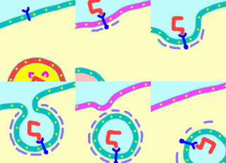
Although there are various processing methods after incorporation, basic mechanisms of endocytosis are almost common. First of all, a receptor ![]() exists. It can not take in anything randomly. Normally, there are various receptors on the cell membrane, but this receptor for endocytosis is a receptor that has the power to distinguish what should be taken into the cell. For the receptor, only the target
exists. It can not take in anything randomly. Normally, there are various receptors on the cell membrane, but this receptor for endocytosis is a receptor that has the power to distinguish what should be taken into the cell. For the receptor, only the target ![]() (mostly seems to be a protein-based one) which is proper for purpose is bound. This receptor is entirely penetrating the cell membrane and as soon as the entrapment object binds to this receptor, the signal is transmitted to the inside of the cell membrane instantaneously. Then a special change occurs inside the cell membrane.
(mostly seems to be a protein-based one) which is proper for purpose is bound. This receptor is entirely penetrating the cell membrane and as soon as the entrapment object binds to this receptor, the signal is transmitted to the inside of the cell membrane instantaneously. Then a special change occurs inside the cell membrane.
● Cell membrane does not stretch or shrink
In the incorporation of a comparatively small object called pinocytosis, the cell membrane of that part begins to collapse when it is bound to a receptor. At first glance, it seems that the cell membrane is stretched and making a bag. Actually, however, unlike rubber balloons, cell membranes are not like pulling and stretching. Phospholipids lined side by side to make a thin sheet. The spacing of phospholipids is not as easy to spread and shrink. So if you try to stretch it by forcibly pulling it will easily be broken. To make a bag, make a dent to make wrinkles little by little from the surroundings to wrap the item with sheets. However, since the components of the cell membrane are quite flowable in the sideways, where it hits the mouth of the bag, unlike the sheet, it becomes smooth and rounded without wrinkle.
● Cell membranes do not make bags themselves
Since the cell membrane itself is only a single sheet of lipid bilayer, it can not do the job of wrapping things in arbitrary way. Therefore, a receiver and a mechanism for it are necessary. It is already written that intracellular changes occur when the subject binds to the receptor.When the receptor reacts, a structure lining the cell membrane is formed inside the cell membrane of that part. ![]() Actin filament grows and spreads if it is phagocytosis which cell membrane is elevated and wrapped around.
Actin filament grows and spreads if it is phagocytosis which cell membrane is elevated and wrapped around.
However, in pinocytosis where the cell membrane falls, special proteins called clathrin are gathered. Clathrin regularly gathers inside the cell membrane and creates an amazing basket structure. While wrapping the cell membrane, the basket draws the whole inside to make a bag of cell membranes.

When a bag like a ball is formed inside the cell, at last, squeeze the narrow mouth formed on the cell surface and cut off the bag from the cell membrane. This is the completion of covered vesicle (coated vesicle). The last scenario is the work of another protein named dynamin (it is omitted in animation). Also, once the vesicles are formed, the basket structure disappears and disappears.
● Vesicles stick to bags again in the cell
The freshly formed vesicles are wrapping the liquid outside the cells, so they are together with the extracellular environment. However, special environments are required to process the entrapped substance. In order to change its environment, it usually fuses and coalesces with another vesicle previously prepared in the cell. This vesicle is called endosome before and after coalescence. Among the endosomes, the environment is stronger than the outside of the cell (pH 6 to 7). As the environment changes, spots naturally separate from the receptors. In many cases, receptors are encapsulated in new vesicles and are incorporated into the cell membrane again and recycled, while the endosomes become more acidic and become called late term endosomes. This is the late stage, the previous stage is the initial endosome.
Further lesion endosomes further incorporate special vesicles (primary lysosomes) ![]() packed with various digestive enzymes. Then, the spots in the endosome are broken down into fragments by strong digestive enzymes (mainly hydrolytic enzymes). Simply referring to lysosomes, it seems to refer to vesicles that wrap digestive enzymes and degradation products together. Among the lysosomes, the acidity is even stronger, which is about pH 5. The degraded product is recycled in the cell as necessary, or discarded outside the cell when it is unnecessary. At last the fate of the object taken from the extracellular endocytosis is over.
packed with various digestive enzymes. Then, the spots in the endosome are broken down into fragments by strong digestive enzymes (mainly hydrolytic enzymes). Simply referring to lysosomes, it seems to refer to vesicles that wrap digestive enzymes and degradation products together. Among the lysosomes, the acidity is even stronger, which is about pH 5. The degraded product is recycled in the cell as necessary, or discarded outside the cell when it is unnecessary. At last the fate of the object taken from the extracellular endocytosis is over.
● Cell membranes need to be supplemented
Since the cell membrane is squeezed as a vesicle every time of endocytosis, the area of the cell membrane becomes smaller by that much, and the volume inside the cell increases because it swallows the extracellular solution. Therefore, if endocytosis is overwhelming, the area of the cell membrane is smaller and smaller, the intracellular volume becomes larger and bigger (?!), surely the cell will rupture. Therefore, at the end of endocytosis, cell membrane replenishment and cellular fluid discharge must be done. In fact, cell membranes are supplemented by vesicles born from within the cell fusing to the cell membrane.

This activity opposite endocytosis is called exocytosis. Exocytosis itself is a phenomenon that has the purpose of secreting the product in the cell and supplying channels and receptors to the cell membrane, in addition to replenishing the cell membrane. At the same time, endocytosis and exocytosis are well-balanced in keeping the cells healthy.
○ Referenced sites
·This is funny! ! ⇒ Anatomical physiology 1 episode “Cellular mechanism” (teacher Tama’s lesson)
○ Related articles
◆[013] Lipid bilayer of the cell membrane ![]()
◆[017] 糖質の吸収 absorption of carbohydratel ![]()
◆[027] ペプチド結合 peptide bond ![]()
◆[007] Glucose and the hydrolysis of sucrose ![]()
◆[019] アデノシン三リン酸(ATP) adenosine triphosphate ![]()
○ Referenced books
・なっとく解剖生理学〈1〉やりとりする細胞と血液 文光堂 (2013/11)
・プロッパー細胞生物学―細胞の基本原理を学ぶ、化学同人
・Essential細胞生物学〈DVD付〉原書第3版、南江堂
・細胞の分子生物学、 ニュートンプレス; 第5版 (2010/01)
・肉単―ギリシャ語・ラテン語 (語源から覚える解剖学英単語集 (筋肉編))
・カラー図解 人体の正常構造と機能 全10巻縮刷版、坂井 建雄、日本医事新報社
・人体機能生理学、杉 晴夫、南江堂
・トートラ人体解剖生理学 原書8版、丸善
・イラスト解剖学、松村 讓兒、中外医学社
・柔道整復学校協会編「生理学」、南江堂
・東洋療法学校協会編「生理学」、医歯薬出版株式会社
rev. 20160205,rev.20170505, rev.20180331.
KISO-IGAKU-KYOIKU-KENKYUKAI(KIKKEN)


